Implant Care and Maintenance
- Dental Health Magazine
- Jul 1, 2020
- 8 min read
Updated: Mar 3, 2021
Source: Dental Health Vol 59 July 2020
Dental implants are long-lasting prosthetic replacements for missing teeth, consisting of:
A titanium screw which is placed into the jawbone
An abutment: an attachment that connects the implant and the implant crown
A crown
In the UK, Bupa estimates that over 130 000 implants are placed per year (1). It is therefore highly important that therapists are well versed in implant assessment and care. There are several evidence-based guidelines that can help the Therapist in approaching implant care:
1. British Society of Periodontology (BSP): focus on peri-implant disease classification
2. Association of Dental Implantology (ADI): focus on all aspects of implant care from examination and diagnosis to treatment and success criteria
3. European Federation of Periodontology (EFP): focus on case definitions and characteristics of peri-implant health and disease
Peri-implant disease
Peri-implant disease is an umbrella term and is caused by bacteria from the oral biofilm. It is essential that we work with the patient to maintain a healthy implant and they should be aware of potential problems.
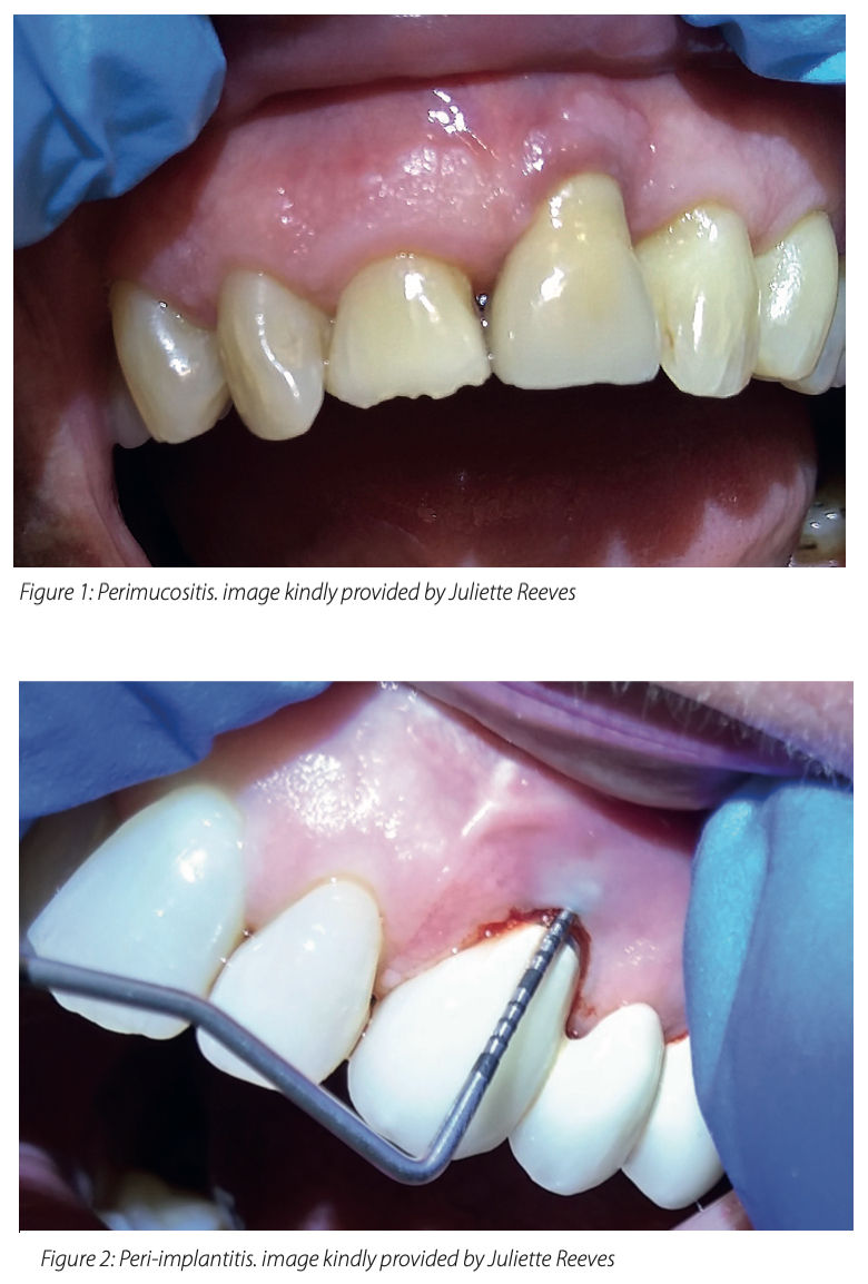
1. Peri-implant mucositis. A reversible inflammatory process in the soft tissues surrounding the implant. Peri-implant mucositis is considered the precursor of peri-implantitis (2).
Figure 1: Peri-implant mucositis
2. Peri-implantitis: This inflammatory process is characterised by the loss of peri-implant bone, increased probing depths, deepening of pockets, clinical signs of inflammation including bleeding on probing and/or pus. Peri-implantitis lesions extend apical of the junctional/ pocket epithelium and are larger than those at peri-implant mucositis and periodontitis sites.
Figure 2: Peri-implantitis
The following are the key points to consider at each stage of the treatment process in a patient who presents with implants:
Implant assessment
Particular attention should always be placed on changes to the previous norm. In new patients, it is advisable, where possible, to ask them to obtain their dental records, including radiographs and pocket charts from their previous implant team to enable you to track and monitor any significant changes.
Obviously, assessment in a maintenance appointment differs from that of an initial examination or re-evaluation appointment. It is useful to ensure that the following have been addressed and recorded in the patient’s notes:
History
Medical history: In particular any changes to current medical status that could affect periodontal status (and thus implant health) e.g. the onset of diabetes
Social history e.g. is there any change in the patient’s smoking status?
Dental history:
The periodontal status of a patient who was previously periodontally stable in remission can deteriorate due to multiple factors including smoking habits, stress, self-care and change in medical status. This can obviously affect implant health
Periodontally affected patients are at higher risk of peri- implantitis.6
Family history: Familial history of periodontal disease can predispose the patient which could affect their implants
Oral hygiene regime: Assess whether the patient is following the suggested routine de rote. If not, it is important to find out what has changed and why?
2. Delivery of smoking cessation advice as appropriate:
Smoking cessation advice and the effects of smoking on the entire dentition including implants should be made clear before implant placement;
The patient should be warned that the failure rate of implant osseointegration is considerably higher among smokers, and maintenance of oral hygiene around the implants and the risk of peri-implantitis are adversely affected by smoking.7
3. Visual assessment of oral hygiene status and health of peri-implant tissues:
To thoroughly check for the presence of plaque and calculus (especially if not plaque disclosing - see below)
To inspect for clinical signs of inflammation
4. Plaque disclosing:
To back up the visual assessment
To use as a tool for patient motivation and education
5. Implant palpation and probing:
Gently running the thumb and forefinger down the length of the implant from root area to crown can reveal any trapped pus or exudate, which might be overlooked on mere visual inspection
Probing can be conducted using a plastic or metal probe to check pocket depths and the presence or absence of bleeding and suppuration
Probing can be difficult due to the bulky nature of implant crowns. This can have a negative effect on correct probing angulation and thus on accurate measurement. Non-removable implant retained dentures can also prove challenging to probe due to their tight placement on the gingivae. When in doubt, gentle probing can be undertaken more than once to ensure accuracy and any difficulties in probing angle or access recorded in the patient’s notes
6. Six-point pocket chart and bleeding score: This should be recorded annually. The patient should have had an initial baseline pocket chart taken when the implant was restored. This initial chart can be used to compare and contrast changes in pocket depth over time and thus influence treatment planning
7. Radiographs: Initial radiographs will have been taken on implant placement. Radiographs are indicated annually and at any point where there are significant changes in your clinical findings. No more than 2mm of bone loss is generally considered acceptable in the first year after placement and 0.2mm in each following year
8. Assessment of implant mobility: This could indicate that the implant crown or screw is loose and not simply failure of osteointegration
It is self-evident that good teamwork and communication with the referring dentist or implantologist is essential. As a minimum, it is advisable that the patient return to the clinician who placed the implant annually to have the implant checked.
Oral Hygiene Instruction
As with all periodontal treatment, prevention is at the heart of everything dental hygienists and therapists do. Moreover, a common patient misconception about implants is that after placement the implant will last a lifetime and does not require any special attention, (or indeed needs less attention than a natural tooth). Oral hygiene instruction and patient motivation are therefore of particular importance. The following tools can help form part of a thorough approach:
Plaque disclosing: Used correctly plaque disclosing is useful in a self-care plan specifically tailored to the patient. It can form a part of both implant assessment and oral hygiene instruction. It is important from the outset that patients with implants understand how important their self-care regime is in keeping their implants healthy
In mouth demonstration: A variety of integral implant care tools are available and should be selected and tailored for each patient. Wherever possible, every effort should be made to demonstrate self-care tools in the patient’s mouth. The patient should be asked to demonstrate back to the clinician what they have been shown to ensure optimal self-care will be achieved at home. It is often only in this demonstration to the clinician that it becomes evident that the patient has not grasped how to use the product
Mouth map: A tailored map of the mouth outlining exactly what products to use where can serve as a useful tool to refer back to. It may be helpful to retain a copy in the notes for reference
Regular checks (in mouth) of self-care tools: This ensures that the best fitting and most appropriate products are being used
Mechanical debridement
Non-surgical periodontal therapy is the primary treatment for peri-implant mucositis. In a longitudinal study of patients diagnosed with peri-implant mucositis, those without adherence to supportive maintenance care yielded a 43.9% incidence of peri-implantitis after five years, compared with 18% in patients adhering to maintenance care.5 This underlines the importance of regular treatment interventions from a dental hygienist or therapist.
However, peri-implantitis can often require surgery and might not respond to simple non-surgical periodontal therapy. As such, an integral part of our role is to work closely with other members of the dental team and refer on as appropriate.
To this end, the ADI maintenance algorithm can prove a useful tool in deciding what treatment approach is most appropriate:

Just like with real teeth, implants can be treated with a number of debridement options:
1. Hand scalers: There is no standard protocol for which exact material implant scalers should be made. However, the following is worthy of consideration:
Titanium coated instruments will not scratch the implant titanium surface.8
Carbon-fibre, teflon and plastic instruments can also be used to remove bacterial deposits. When thin, scalers made of these materials tend to break easily and when thick they can often prove too bulky for effective use. It has been found that plastic instruments have no effect on implant surfaces but they do leave plastic debris which could not be removed by a jet of air commonly used to clean surfaces for SEM. This debris could also provide additional pockets of a size suitable for bacterial colonization.9
Steel instruments are not favoured by many as they have an external hardness higher than titanium and thus can cause scratches.10

2. Ultrasonic scalers: In particular, polyetheretherketone-coated tips serve to debride the implant surface and afford patient comfort.
3. Air polishing systems: In contrast to sodium bicarbonate air spray which is used for polishing, amino-acid glycine powder can be used for biofilm disruption supra and subgingivally without damaging the implant surface.
4. Polishing: A rubber cup and implant safe low abrasive polishing paste can be used on the implant crown.
Setting recall interval
Recall intervals should be determined by patient susceptibility and need: Upon implant placement adequate time should be left for healing before any mechanical debridement is undertaken. Hygiene appointments should focus on oral hygiene instruction during this time. You should liaise with the implantologist to determine when mechanical debridement should commence. A three-month time frame post placement is often favoured.
As with natural teeth, risk factors should be considered when deciding the recall interval (e.g. medical, social, family history).
Recall frequency is best set using longitudinal information. It is therefore advisable that the patient attend more frequently (for instance every three months) in the first year after implant placement. If the patient has an adequate level of plaque control and the comparison of present with past measurements indicates stability, the interval can be prolonged up to six months by increments of one month from visit to visit.
Conclusion
For many implant patients, the dental hygienist or therapist is the key point of contact in the practice, often seeing implant patients more than other team members. As such, an integral part of our role is identifying changes to implants and referring cases on to other team members in a timely fashion.
There are several evidence-based guidelines available in the classification, assessment, maintenance and recall setting for patients with peri-implant disease. In particular the Association of Dental Implantology guide can form good practice recommendations. The whole implant team within an individual practice can benefit from establishing practice protocols for patient treatment using up to date best practice information and research.
References
1. https://www.bupa.co.uk/dental/dental-care/treatments/implants- dentures-bridges/dental-implants 2. British Society of Periodontology. New classification of periodontal and peri-implant diseases and conditions. 2018. 3. Ucer, C; Wright, S; Scher E et al. Association of Dental Implantology Guidelines on Peri-implant Monitoring and Maintenance. 4. Caton JG, Armitage G, Berglundh T et al. A new classification scheme for periodontal and peri-implant diseases and conditions – Introduction and key changes from the 1999 classification. J Periodontol. 2018;89:S1-8. 5. Costa FO, Takenaka-Martinez S, Cota LOO et al. Peri-implant disease in subjects with and without preventive maintenance: a 5-year follow-up. J Clin Periodontol. 2012;39(2):173-181. 6. Heitz-Mayfield LJA. Peri-implant diseases: Diagnosis and risk indicators. J Clin Periodontol. 2008;35(8 Suppl):292-304 7. Lambert PM, Morris HF, Ochi S. The influence of smoking on 3-year clinical success of osseointegrated dental implants. Ann Periodontol. 2000; 5(1):79–89. 8. Lang NP, Salvi GE, Sculean A. Nonsurgical therapy for teeth and implants – when and why? Periodontol. 2000. 2019;79(1):15-21. 9. Driver J. SEM Study Comparing Instrumentation Result/Observations. University of Montana Division of Biological Science, 2009. 10. Mombelli A. Maintenance therapy for teeth and implants. Periodontol 2000. 2019; 79(1):190-199


Comments